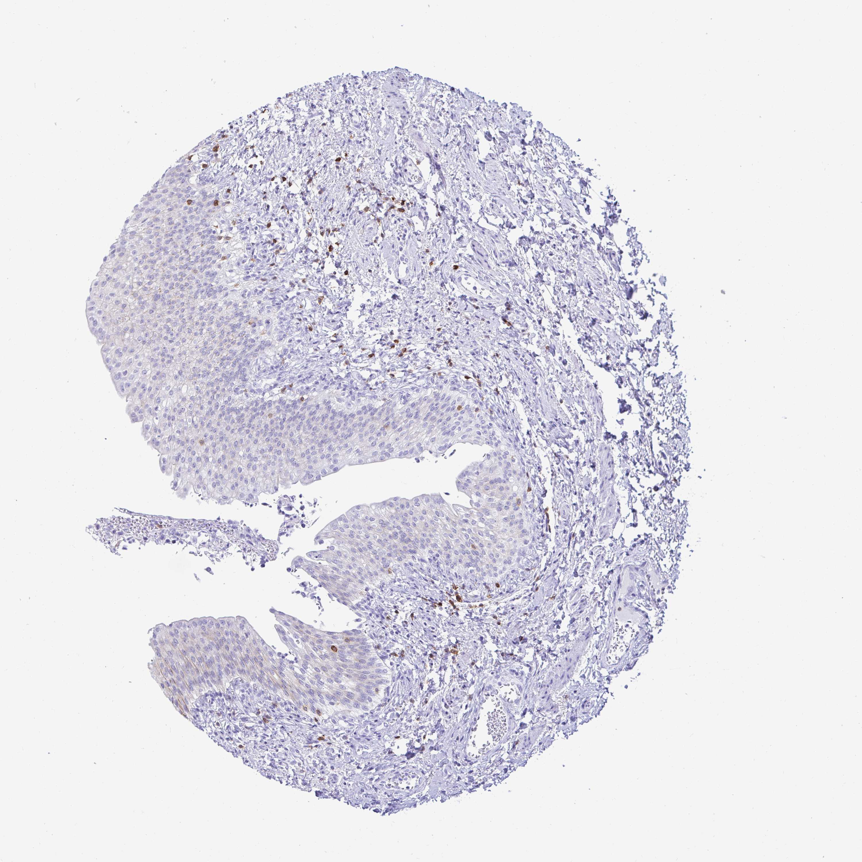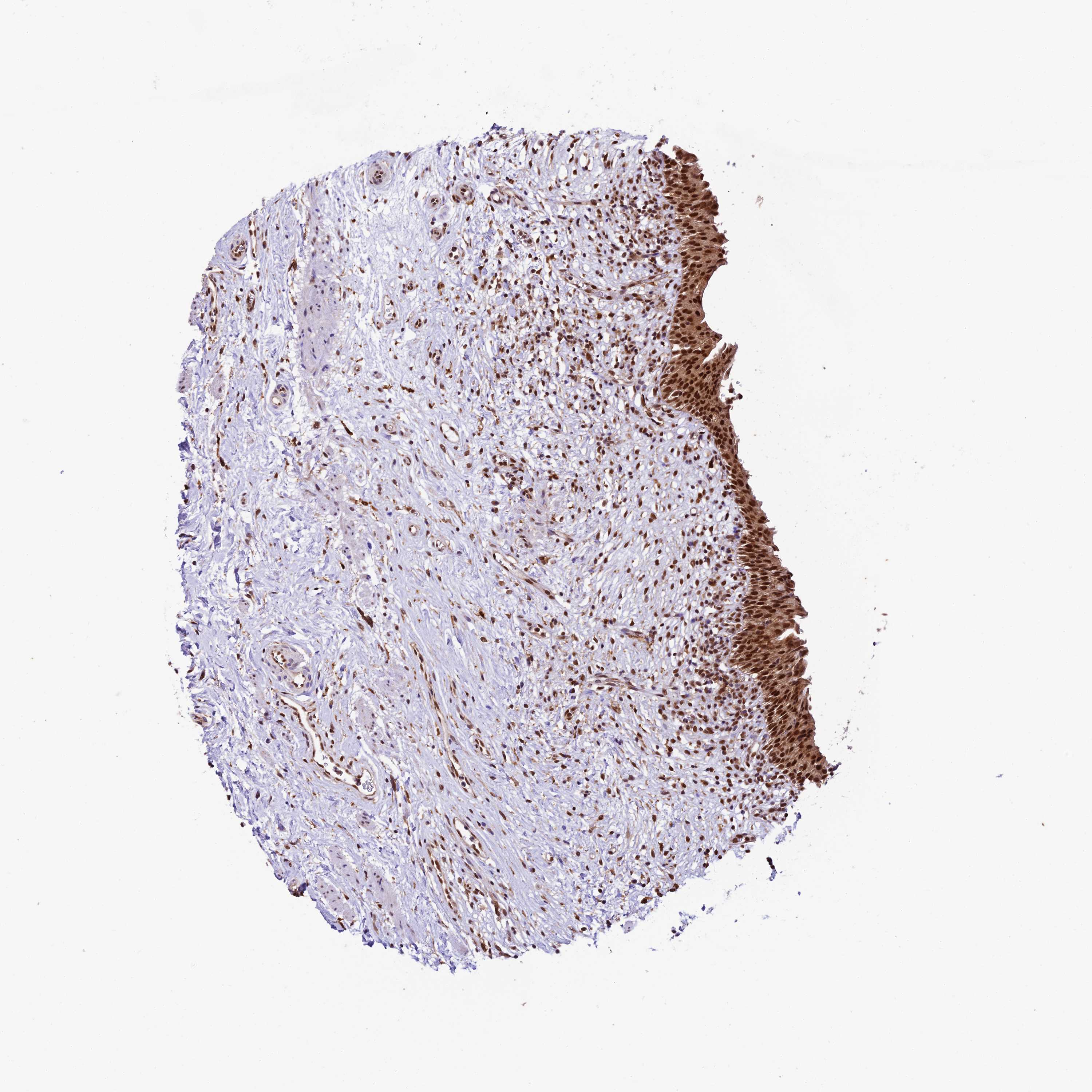
Therefore, the aim of this retrospective study is to evaluate voided urine samples reported as atypical and to assess the clinical significance of this category through histologic correlation of these samples.


Thus, in the absence of agreement and the lack of diagnostic criteria for urothelial atypia, the atypical urothelial cell category remains one of the challenging diagnostic entities. However, the criteria to separate reactive from neoplastic atypia are not well defined in this article or in the literature, in general. In 2004, the Papanicolaoau Society of Cytopatholgy recommended to include “atypical urothelial cells” as a diagnostic category in the urine cytology, with a comment in the report to further classify the atypia as reactive or neoplastic. However, there is lack of consensus regarding the terminology and the diagnostic criteria that should be used for urothelial atypia and the “atypical” category remains a wastebasket diagnosis that is used variably by individual cytopathologists in different institutions. These cases often fall into the atypical categories. There are several situations that can affect the cellularity and the cytology of the cells, including instrumentation, inflammation, infection, surgical manipulation, treatment with chemo and radiotherapy and calculi, making it difficult even for the experts to reliably discriminate malignant cells. Finally, it has been shown by several studies that increasing the number of the samples will increase the sensitivity of urine cytology, especially for the detections of high-grade lesions. This in fact could be explained by the absence of the instrumentation-induced reactive changes. In addition, specimen type can also affect the interpretation of urine cytology, with voided specimens being more specific but slightly less sensitive than instrumented urine. However, low-grade tumors are not detected reliably by cytology, with sensitivity and specificity values as low as 8.5 and 50%, respectively. It has been widely accepted for the diagnosis of high-grade urothelial carcinoma with a sensitivity as high as 98%. The accuracy of urine cytology diagnosis depends on several factors that are related to tumor grade, type of the specimen and sampling. The second indication is follow-up of patient with bladder cancer and third is as screening of high-risk groups for bladder cancer such as those exposed to aniline dye or to aromatic amines and those with history of urinary bilharziasis.

Indications for urine cytology fall mainly into three categories the most common one is patients with hematuria. The examination of urine sediment smears was first popularized by George Pananicolaou and Marshall in the 1940s for bladder cancer detection and follow up. Urine examination is considered to be one of the oldest clinical laboratory tests known to humans.


 0 kommentar(er)
